Alessandro Mancini and Luca De Carolis are Co-first authors.
Nicodemo Baffa, Brunangelo Falini and Enrico Tiacci are Co-last authors.
INTRODUCTION
HCL is an indolent BRAF-mutated leukemia usually presenting with cytopenias, splenomegaly, little or no lymphadenopathy, heavy bone marrow (BM) infiltration and few circulating leukemic cells. Imaging exams for staging include abdominal echography and computed tomography (CT), while the role of PET is largely unknown.
METHODS
84 patients (pts) with relapsed/refractory HCL participating to our clinical trials of BRAF inhibitor-based therapies prospectively underwent PET/CT pre- and post-treatment, in addition to usual examinations for response assessment. Since FDG uptake is normally higher in liver than spleen, BM and lymph nodes, uptake in the latter tissues was considered abnormal if equal to or higher than the liver. Splenomegaly was defined by a longest spleen diameter >13 cm on PET/CT. The standard definition of complete remission (CR) in HCL just requires no palpable splenomegaly on physical examination, in addition to resolution of cytopenias as well as no hairy cells visible in the blood smear and the BM biopsy by non-immunological stains.
RESULTS
Pre-therapy, 58/77 (75%) non-splenectomized pts showed abnormal splenic FDG uptake (maximal standardized uptake value/SUVmax: median 4.8, vs 3.9 for the liver), which was always diffuse without focal lesions. Conversely, focal splenic uptake is not infrequent in splenic marginal zone lymphoma (18%, 7/39 pts - Abdom Radiol 2018;43:2721; vs of 0/58 HCL pts, p=0.0003), which may aid in the differential diagnosis with this HCL-mimicker. Moreover, 14/58 (24%) HCL pts. with a PET+ spleen did not have splenomegaly, and splenic uptake returned to normal in 10/11 evaluable cases (91%) achieving at least a partial response post-therapy, which suggests leukemic spleen involvement even without splenomegaly.
Abnormal diffuse BM uptake was observed in 41/82 evaluable pts (50%; median SUVmax 5, vs 3.4 for the liver). In these cases, leukemic involvement in the BM biopsy was greater (median 80%) than in the other 41 pts (median 65%; p-value <0.01), pointing to HCL infiltration as the cause of abnormal BM uptake rather than to reactive hyperplasia of non-involved BM.
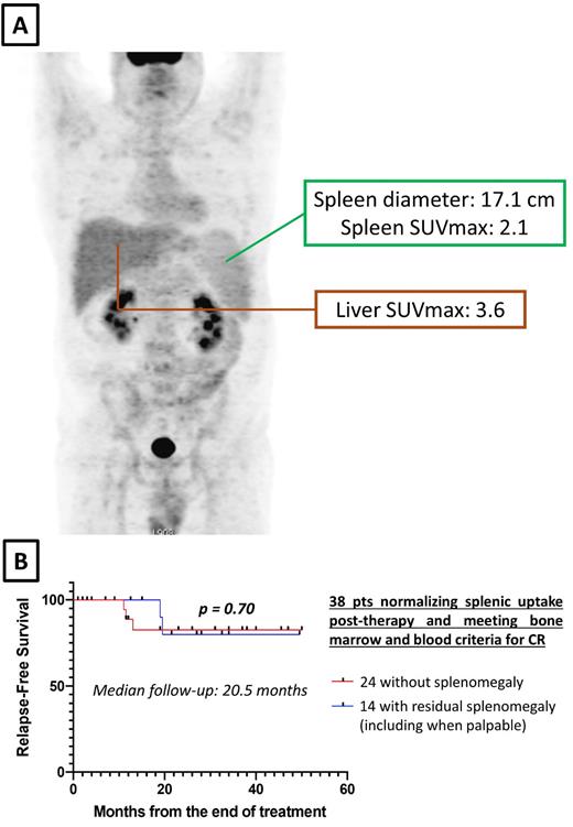
Post-therapy, pathologic spleen uptake normalized in 46/51 evaluable pts (90%), and their CR rate was higher (32/46 pts, 70%) than in the other 5 pts remaining PET+ (1/5, 20%; p=0.047). Among the 46 PET- cases, 31 (67%) were not splenomegalic while 15 (33%) had residual splenomegaly (up to 17 cm; Fig. 1A); interestingly, within PET- cases relapse-free survival (RFS) was similar not only in conventionally defined CR cases with (n=8) vs without (n=24) residual non-palpable splenomegaly (p=0.84 after a median follow-up of 19 months), but also when the 6 cases meeting all conventional CR criteria except for still palpable splenomegaly were added to the 8 CR pts with residual non-palpable splenomegaly (p=0.7 after a median follow-up of 20.5 months - Fig. 1B). These findings suggest that metabolic status reflects splenic HCL involvement more reliably than spleen size or palpability, a concept that could improve the current definition of CR in HCL.
Abnormal BM uptake resolved after treatment in 34/37 evaluable pts (92%), and their CR rate was again higher (23/34, 68%) than in the other 3 pts remaining PET+ (0/3; p=0.047). Leukemic infiltration of the BM biopsy was lower in the 34 PET- vs the 3 PET+ pts (median 5% vs 70%-80%, respectively; p=0.0004), indicating that BM abnormal uptake reflects HCL infiltration load rather than reactive BM hyperplasia post-therapy.
Finally, abnormal lymph node uptake was observed pre-therapy in 9/84 (11%) pts (median SUVmax 7.1, vs 3.4 for the liver) and normalized after treatment in 7/8 evaluable pts (88%), including one case with residual lymph node enlargement (2.2 cm); all these 7 pts had achieved a CR while the remaining pt had no response to therapy.
CONCLUSIONS
PET is potentially useful in the differential diagnosis with splenic marginal zone lymphoma and may aid in the clinical management of HCL pts by detecting metabolic involvement of the main disease sites even in the absence of organomegaly. Importantly, PET signal tracks with response to therapy even in case of persistent organomegaly, which may lead to refine the definition of CR in HCL.
OffLabel Disclosure:
Zaja:Sobi: Honoraria, Research Funding; Grifols: Consultancy, Honoraria, Research Funding; Amgen: Honoraria, Research Funding; Novartis: Consultancy, Honoraria, Membership on an entity's Board of Directors or advisory committees, Research Funding. Tiacci:Kite-Gilead: Consultancy; Deciphera: Consultancy; Innate Pharma: Consultancy.
This presentation include information regarding vemurafenib (BRAF inhibitor), cobimetinib (MEK inhibitor) and obinutuzumab (anti-CD20 antibody) in the context of Hairy Cell leukemia.


